Showing posts with label Health. Show all posts

Disaster’s Hidden Toll


 image +

Credit NHK News – Fukushima evacuation zone March 2011


# 6846


With our short attention span, and the news media’s proclivity for moving on to the next big disaster or story, we often don’t closely follow the struggle to rebuild disaster stricken communities, which can take months or even years.


And for some caught in harm’s way, whose homes and businesses were destroyed - and loved ones lost - there is no going back to the way things used to be.


Not surprisingly, that can produce significant mental and physical health challenges for those affected.


Today we’ve a report out of Japan showing that the earthquake/tsunami of March 2011 that killed more than 20,000 people in Japan also had a long-term, largely unseen, effect on nursing home patients who were forced to evacuate to temporary facilities.


A study shows a 2.4 fold increase in deaths during the 8 months following the earthquake.  Deaths not caused by the quake, tsunami, or radiation release itself – but likely brought on by the stress of having to live in make-shift emergency shelters.


A unusually large number of these excess deaths were due to pneumonia or bronchitis, which many attribute to insufficient emergency shelters provided for the elderly and frail.


This report from the Ashasi Simbum.


Death rates spike among elderly evacuees from Fukushima

January 11, 2013

By YURI OIWA/ Staff Writer

Former residents of nursing homes near the Fukushima No. 1 nuclear plant died at a higher rate than usual in 2011, a study has shown, likely because of the stress of evacuation and having to live in temporary accommodations such as draughty school gyms.


Researchers from the Fukushima Medical University studied reports submitted to the Fukushima prefectural government by 34 institutions for the elderly and found that the death rate over eight months in 2011 was 2.4 times that of the same period in 2010.

(Continue . . . )


A similar result was found in this study of nursing home evacuations from the University of South Florida.


The Effects of Evacuation on Nursing Home Residents With Dementia

Lisa M. Brown, PhD, David M. Dosa, MD, MPH, Kali Thomas, MA, Kathryn Hyer, PhD, MPP, Zhanlian Feng, PhD, Vincent Mor, PhD

Background: In response to the hurricane-related deaths of nursing home residents, there has been a steady increase in the number of facilities that evacuate under storm threat. This study examined the effects of evacuation during Hurricane Gustav on residents who were cognitively impaired.


Conclusions: The findings of this research reveal the deleterious effects of evacuation on residents with severe dementia. Interventions need to be developed and tested to determine the best methods for protecting this at risk population when there are no other options than to evacuate the facility.


We’ve looked at other post-disaster health impacts in the past, such as in Post Disaster Stress & Suicide Rates. One disaster discussed was a 1999 7.3 earthquake that struck in Chi-Chi, Nantou county in central Taiwan killing more than 2,300 people.


A study that subsequently appeared in the Taiwan Journal of Medicine (Disease-specific Mortality Associated with Earthquake in Taiwan Hsien-Wen Kuo, Shu-Jen Wu, Ming-Chu Chiu) found `a considerable increase in the number of suicides after the earthquake’.


PTSD (Post Traumatic Stress Disorder) can often occur in the wake of a disaster or traumatic experience. Symptoms may include anxiety, depression, suicide and PTSD may even lead to drug and alcohol-related disorders.


Victims of personal violence, rescue and medical workers, victims of disasters, terrorism, physical or psychological trauma, and/or a combat zone are all at risk of suffering some level of PTSD.


In Psychological First Aid: The WHO Guide For Field Workers we looked at the need for, and a guide for providing psychological first aid (PFA) in a post-disaster environment.


The CDC also provides a website which contains a number of resources devoted to coping with disasters.


Coping With a Disaster or Traumatic Event

Trauma and Disaster Mental Health Resources

The effects of a disaster, terrorist attack, or other public health emergency can be long-lasting, and the resulting trauma can reverberate even with those not directly affected by the disaster. This page provides general strategies for promoting mental health and resilience. These strategies were developed by various organizations based on experiences in prior disasters.


As does the National Center For PTSD - including videos - on how to provide Psychological First Aid.

A reminder that a disaster’s impact can linger long after the story has fallen off the front pages, and that indirect casualties can follow months after the initial event.

»» Read More

PLoS One: Influenza Viral Shedding & Asymptomatic Infections

image

Photo Credit PHIL (Public Health Image Library)


# 6776


Despite major advances in the study of influenza viruses, there remain significant gaps in our understanding of just how they work once they infect a human (or any other animal) host. 


Basic questions, such as `How long are we infectious?, or `How common are asymptomatic infections?  remain only partially answered.


Complicating matters, variations in individual host’s immune responses, and different strains of flu may produce varying results. Meaning that most studies can only add incrementally to our knowledge, rather than completely answering these questions.


Previously, we’ve seen evidence of asymptomatic and `presymptomatic’ shedding of influenza viruses.


In 2011, in EID Journal: Pre-Symptomatic Influenza Transmission we looked at three clusters of suspected pre-symptomatic transmission of the 2009 H1N1 virus in Japan.


And in Pre-Symptomatic Transmission Of H1N1 Influenza In the Ferret Model, researchers inoculated ferrets with the 2009 H1N1 flu, and then placed them near uninfected ferrets (some in direct contact, others in adjacent cages) at different stages after infection.


They then tested the exposed ferrets to see when, and under what circumstances, they became infected. They found that ferrets became infectious just 24 hours after becoming infected, and nearly 24 hours before showing the earliest outward signs of infection (fever).


The importance of all of this is, if presymptomatic and asymptomatic carriers of a flu virus are able to efficiently transmit the illness on to others, then strategies that seek to identify and isolate flu cases would have only limited success in containing a pandemic.


Similarly, understanding how long a person sheds the virus after becoming infected is crucial, so we can know when it is (relatively) safe for flu victims to return to work or school without endangering others.


The CDC’s general take on this topic is:


The Flu Is Contagious

Most healthy adults may be able to infect others beginning 1 day before symptoms develop and up to 5 to 7 days after becoming sick. Children may pass the virus for longer than 7 days. Symptoms start 1 to 4 days after the virus enters the body. That means that you may be able to pass on the flu to someone else before you know you are sick, as well as while you are sick. Some persons can be infected with the flu virus but have no symptoms. During this time, those persons may still spread the virus to others.


Yesterday, a new study appeared in PloS One, conducted in Germany over 4 flu seasons (2007-2011) and involving 4 flu strains - seasonal (A(H3N2), A(H1N1), influenza B, and pandemic (A(H1N1)pdm09 - that looks at many of these transmission issues. 


Comparison of Shedding Characteristics of Seasonal Influenza Virus (Sub)Types and Influenza A(H1N1)pdm09; Germany, 2007–2011

Thorsten Suess, Cornelius Remschmidt, Susanne B. Schink, Brunhilde Schweiger, Alla Heider, Jeanette Milde, Andreas Nitsche, Kati Schroeder, Joerg Doellinger, Christian Braun, Walter Haas, Gérard Krause, Udo Buchholz

Background

Influenza viral shedding studies provide fundamental information for preventive strategies and modelling exercises. We conducted a prospective household study to investigate viral shedding in seasonal and pandemic influenza between 2007 and 2011 in Berlin and Munich, Germany.

Methods

Study physicians recruited index patients and their household members. Serial nasal specimens were obtained from all household members over at least eight days and tested quantitatively by qRT-PCR for the influenza virus (sub)type of the index patient. A subset of samples was also tested by viral culture. Symptoms were recorded daily.

Results

We recruited 122 index patients and 320 household contacts, of which 67 became secondary household cases. Among all 189 influenza cases, 12 were infected with seasonal/prepandemic influenza A(H1N1), 19 with A(H3N2), 60 with influenza B, and 98 with A(H1N1)pdm09. Nine (14%) of 65 non-vaccinated secondary cases were asymptomatic/subclinical (0 (0%) of 21 children, 9 (21%) of 44 adults; p = 0.03).


Viral load among patients with influenza-like illness (ILI) peaked on illness days 1, 2 or 3 for all (sub)types and declined steadily until days 7–9. Clinical symptom scores roughly paralleled viral shedding dynamics.


On the first day prior to symptom onset 30% (12/40) of specimens were positive. Viral load in 6 asymptomatic/subclinical patients was similar to that in ILI-patients. Duration of infectiousness as measured by viral culture lasted approximately until illness days 4–6. Viral load did not seem to be influenced by antiviral therapy, age or vaccination status.

Conclusion

Asymptomatic/subclinical infections occur infrequently, but may be associated with substantial amounts of viral shedding. Presymptomatic shedding may arise in one third of cases, and shedding characteristics appear to be independent of (seasonal or pandemic) (sub)type, age, antiviral therapy or vaccination; however the power to find moderate differences was limited.


While this was a relatively small study, and their findings don’t always align perfectly with others we’ve seen (for instance, children didn’t appear contagious any longer than adults), it does provide us with some interesting data.


  • First, nearly 1/3rd of cases began shedding virus while pre-symptomatic
  • Second, viral loads in (six studied) asymptomatic cases were similar to that to patients exhibiting ILI (influenza-like-illness) symptoms.
  • Third, viral load among symptomatic patients peaked on illness days 1, 2 or 3 and declined steadily until days 7–9


Some other gems (bolding mine) excerpted from this open access article include:


  • Overall 63% of non-vaccinated secondary household cases had an ILI-syndrome and the proportion of asymptomatic/subclinical secondary cases was 14%.
  • Frequency distribution of clinical symptoms did not differ between A(H1N1)pdm09 cases and non-pandemic influenza cases.
  • Interestingly, 21% of adult secondary cases were asymptomatic/subclinical, while all children that contracted influenza were symptomatic.
  • Based on the population of ambulatory patients investigated we found no evidence that the amount of shedding is particularly higher in children, nor that duration of viral shedding is significantly longer in children compared to adults.


The authors conclude by saying:


In summary, our study addresses several important questions on clinical manifestation, duration of infectiousness, viral shedding patterns, including shedding before symptom onset and in asymptomatic/subclinical patients, as well as the effect of vaccination and antiviral therapy on viral shedding.


Important single results include the finding that children do not seem to be infected asymptomatically, that shedding one day before symptom onset may occur in one third of influenza patients, that asymptomatic/subclinical influenza patients occur rarely, but viral load (and probably infectiousness) may be substantial, and vaccinated influenza patients do not show different shedding patterns compared to non-vaccinated cases with ILI.


Overall results do not show marked differences between seasonal influenza (sub)types and influenza A(H1N1)pdm09.

»» Read More

Early Flu Cases Begin To Emerge

 image

Photo Credit CDC Influenza Home Care Guide

# 6745


While it may not tell us a lot about how the rest of the 2012-2013 flu season will go, over the past couple of weeks several states have begun reporting spikes in early influenza activity.  The last FluView report from the CDC (Nov 17th) indicated flu activity was increasing in parts of the country; notably in the south central and southeastern states.


Likewise, the Flu Near You weekly online survey (of self reported symptoms) shows the greatest rate of ILI (Influenza-like-Illness) activity currently in the middle southern states.


image

Flu Near You map  11/29/12


NOTE: ILI’s can include many non-influenza viral illnesses, including adenovirus, parainfluenza, rhinovirus and others which are indistinguishable from influenza without laboratory tests (see Dozens Of Ways To Spell `I-L-I’).


Although `flu season’ can begin as early as October some years, it is generally December before the virus really begins to make its presence widely known. The Thanksgiving holiday, which often brings many family members together, may play a part in kick starting the epidemic each year.


Yesterday South Carolina’s Department of Health and Environmental Control issued the following notice, which included word of a pediatric flu fatality.


FOR IMMEDIATE RELEASE
Nov. 28, 2012

Flu cases spiking early, first flu death in South Carolina

COLUMBIA, S.C. – The S.C. Department of Health and Environmental Control notes the state’s first flu-associated death of the season, as well as a significant and earlier-than-normal increase in influenza activity, the agency announced today.


“Tragically, a child from Barnwell County has become our first confirmed influenza-associated death of the season,” said Linda Bell, M.D. and interim state epidemiologist. “The flu can be especially serious for the very young and the elderly.


“Our latest statewide activity report indicates that influenza has quickly reached ‘widespread’ levels in South Carolina,” Dr. Bell said. “Flu activity typically peaks in February, and it is very unusual for us to see this number of cases so early in the season. Therefore, we strongly encourage vaccination to prevent the flu and its potentially serious consequences.”

(Continue . . .)


Similarly, reports from Central Florida indicate an early start here as well.  This from the Orlando Sentinel.


Flu season hitting earlier, local clinics say

1:09 p.m. EST, November 27, 2012|By Marni Jameson, Orlando Sentinel

Flu season has arrived in Central Florida, and it's well ahead of schedule, according to Dr. Tim Hendrix, medical director for CentraCare, which operates 21 clinics throughout Central Florida.


The CentraCare clinics saw 250 confirmed cases of the flu last week alone. That's more than a 10-fold increase in flu cases compared to Thanksgiving week last year, when the clinics reported 21 confirmed cases, said Hendrix.

(Continue . . .)


Other states now confirming flu activity include Arizona, Missouri, Colorado, Maine, New York and Ohio. Most surveillance reports are trailing indicators – showing us the level of activity 1 to  2 weeks ago – so the level of activity today could be different.


In any event, if you haven’t gotten your flu shot, now would be an excellent time to do so, as it takes a couple of weeks to begin building antibodies once you get the shot.


No, it won’t protect you against non-influenza viral illnesses, and the protection it provides against the flu can vary from year-to-year and person-to-person. 


A meta-analysis by CIDRAP in 2011 (see A Comprehensive Flu Vaccine Effectiveness Meta-Analysis) found the trivalent inactivated vaccine (TIV) had a combined efficacy of 59% among healthy adults (aged 18–65 years).


Still, flu shots have an excellent safety profile and remain one of the most effective preventatives against catching influenza. Beyond that, being vigilant (read: obsessive) regarding day-to-day flu hygiene is your best safeguard.


The CDC recommends:


Take everyday preventive actions to stop the spread of germs.

  • Cover your nose and mouth with a tissue when you cough or sneeze. Throw the tissue in the trash after you use it.
  • Wash your hands often with soap and water. If soap and water are not available, use an alcohol-based hand rub.
  • Avoid touching your eyes, nose and mouth. Germs spread this way.
  • Try to avoid close contact with sick people.
  • If you are sick with flu-like illness, CDC recommends that you stay home for at least 24 hours after your fever is gone except to get medical care or for other necessities. (Your fever should be gone without the use of a fever-reducing medicine.)
  • While sick, limit contact with others as much as possible to keep from infecting them.
  • See Everyday Preventive Actions  [257 KB, 2 pages] and Nonpharmaceutical Interventions (NPIs) for more information about actions, apart from getting vaccinated and taking medicine, that people and communities can take to help slow the spread of illnesses like influenza (flu).


Of course, if all of these preventatives fail, stay home so you don’t share your virus with the world. If you are at high risk of complications, contact your doctor to see about taking antiviral medications.

image

Otherwise, CDC’s Influenza Home Care Guide should see you through.

»» Read More

Hong Kong’s Coronavirus Response

image

Coronavirus – Credit CDC PHIL


# 6738


No city experienced a greater impact from the SARS epidemic a decade ago than did Hong Kong in 2003. Between March 11th and June 6th, a total of 1750 cases were identified, and of those, 286 died.


From the JRSM (Journal of the The Royal Society of Medicine) in August of 2003, we get this description of the spread of the disease in the city.


 The SARS epidemic in Hong Kong: what lessons have we learned?)

Lee Shiu Hung, MD FFCM

(Excerpt)

The SARS epidemic in Hong Kong has gone through three phases. The first was an explosive outbreak in a teaching hospital, affecting a large number of hospital staff and medical students. This phase took place in March 2003.

The second phase was an outbreak in the community as a result of the spread of infection from the hospital to the community. This reached its peak in early April 2003 when the disease affected a housing estate known as Amoy Gardens; a total of 329 residents in that estate came down with the disease and 33 died.

The third phase began in early May, with continuing occurrence of the disease in eight hospitals and more than 170 housing estates throughout the city but with the daily number of new cases declining from double to single digits in mid-June (the time of writing).

(Continue . . .)


Out of this trial by fire, Hong Kong’s Centre For Health Protection was born, and today it arguably runs one of the most proactive disease surveillance systems in the world.

So one should not be surprised to find a link to the new coronavirus front and center on the CHP’s home page.

image


On September 27th, just 3 days after being notified of the first coronavirus case, Hong Kong modified their Prevention and Control of Disease Ordinance (Cap. 599) to include “Severe Respiratory Disease associated with Novel Coronavirus”  as one of the statutorily notifiable diseases (see Letter To Doctors).


You’ll find a variety of notices, and letters to institutions regarding this emergent coronavirus on this page.


Today the Hong Kong government posted some remarks by their Secretary for Food and Health, Dr Ko Wing-man. As you’ll see, that while they are watching this situation carefully, at this time they have no plans to upgrade their alert level.


SFH on overseas cases of Severe Respiratory Disease associated with Novel Coronavirus


Following is the transcript of remarks made by the Secretary for Food and Health, Dr Ko Wing-man, after attending a public function this afternoon (November 25):

Secretary for Food and Health: Regarding the new novel coronavirus causing severe respiratory disease, there is a concern that over a span of two months, another batch of four new cases (three from Kingdom of Saudi Arabia and one from Qatar) was confirmed by the World Health Organization (WHO). The WHO also pointed out that we cannot assume the source of infection is only present in the countries concerned. Patients who have not got a history of travel to these two particular middle-east countries but developed symptoms of illness similar to the diseases caused by this new novel coronavirus, they might still be considered necessary to undertake the test for the new coronavirus.

Reporter: (On emergency level in Hong Kong for an outbreak of the new disease)

Secretary for Food and Health: The Centre for Health Protection of the Department of Health is consistently conducting risk assessment, following up on whether there will be any new information or new cases reported. Up to this moment, there is no need to upgrade our response level for the infectious disease outbreak. However, the new reported cases (due to infection with the novel coronavirus) highlighted the need to enhance our surveillance measures, both at the hospitals as well as at the immigration check points. 

(Please also refer to the Chinese portion of the transcript.)

Ends/Sunday, November 25, 2012
Issued at HKT 20:23


You can pretty much expect an enhanced level of surveillance around the world in the coming days as a result of last week’s announcement from the World Health Organization (see WHO Announces Additional Coronavirus Cases).


It is far too soon to know whether this virus will pose a major public health threat, but one of the lessons learned from the SARS outbreak of 2003 is that delays in reporting can have deadly consequences.


It is far better for public health officials to be hyper-vigilant today, than to have to play catch up tomorrow.

»» Read More

Studies Weigh In On The `Obesity Paradox’


image

Photo Credit CDC PHIL


# 6651


While just about everyone will concede that being lean and athletic is preferable to being soft and pudgy, there is a growing body of evidence that – with some medical conditions, at least - having a bit of extra poundage could be beneficial to your long-term survival. 


Research has shown that patients having a BMI > 30 actually are more likely to survive certain medical conditions than patients with a normal or below-normal body mass index.


It’s called the `Obesity Paradox’, and quite frankly, it’s been driving doctors and researchers just a little bit nuts for years.  


Now, before anyone cries `foul’, there are plenty of health risks that come from being obese, including diabetes, coronary artery disease, sleep apena, hypertension, and stroke. The point here isn’t that being overweight is healthier.


it isn’t.


But for people with certain medical conditions, including diabetes, congestive heart failure, kidney dialysis, heart attacks, and Asthma - carrying extra pounds appears to improve their outcomes.


This curious (and controversial) finding was first described in the literature in 1999, regarding survival rates of patients on kidney dialysis (see Influence of excess weight on mortality and hospital stay in 1346 hemodialysis patients). 


Since then, this phenomenon has inspired a good deal of research, much of it coming to similar conclusions.


Overnight we saw this press release come from the American College of Chest Physicians.


'Obesity paradox': Extra weight linked to better outcomes for septic shock, asthma exacerbation

Although obesity is linked to a variety of health risks, new research indicates that obese patients may have an advantage over nonobese patients in certain health situations, including septic shock and acute asthma exacerbation.


In two separate studies presented at CHEST 2012, the annual meeting of the American College of Chest Physicians, researchers compared outcomes in obese (BMI >30) vs nonobese patients with either septic shock or acute asthma exacerbation. Results showed that, although obese patients with asthma are more at risk for asthma exacerbations, near fatal exacerbations were more prevalent in nonobese patients.


Likewise, obese patients with septic shock had decreased mortality compared with nonobese patients. Researchers attribute this "obesity paradox" partly to a blunted pro-inflammatory cytokine response in obese patients.


Recently,  JAMA published a pooled analysis of 5 cohort studies that found – surprisingly – that : “Adults who were normal weight at the time of incident diabetes had higher mortality than adults who are overweight or obese.”

Original Contribution | August 8, 2012

Association of Weight Status With Mortality in Adults With Incident Diabetes

Mercedes R. Carnethon, PhD; Peter John D. De Chavez, MS; Mary L. Biggs, PhD; Cora E. Lewis, MD; James S. Pankow, PhD; Alain G. Bertoni, MD, MS; Sherita H. Golden, MD, MS; Kiang Liu, PhD; Kenneth J. Mukamal, MD, MPH; Brenda Campbell-Jenkins, PhD; Alan R. Dyer, PhD

JAMA. 2012;308(6):581-590. doi:10.1001/jama.2012.9282.


We’ve another large study out of Sweden that looked at patient outcomes with acute coronary syndromes (ACSs)  – and you guessed it – obese patients had better survival rates after a heart attack than patients of normal weight.


Evidence for obesity paradox in patients with acute coronary syndromes: a report from the Swedish Coronary Angiography and Angioplasty Registry

Oskar Angerås, Per Albertsson, Kristjan Karason, Truls Råmunddal, Göran Matejka, Stefan James, Bo Lagerqvist, Annika Rosengren and Elmir Omerovic

Conclusion In this large and unselected group of patients with ACSs, the relation between BMI and mortality was U-shaped, with the nadir among overweight or obese patients and underweight and normal-weight patients having the highest risk. These data strengthen the concept of the obesity paradox substantially.


Similarly, from the American Journal of Cardiology, we get this study from earlier this summer, on survival rates of patients with congestive heart failure.


Volume 110, Issue 1 , Pages 77-82, 1 July 2012

The Obesity Paradox in Men Versus Women With Systolic Heart Failure

Adrienne L. Clark, BA, Jennifer Chyu, Tamara B. Horwich, MD, MS

Abstract (excerpt):

In multivariate analyses, normal BMI and normal WC were associated with higher relative risk for the primary outcome in men (BMI 1.34, WC 2.02) and women (BMI 1.38, WC 2.99). In conclusion, in patients with advanced HF, high BMI and WC were associated with improved outcomes in both genders. Further investigation of the interaction between body composition and gender in HF outcomes is warranted.


Admittedly, many of these medical conditions may well have been brought on by the patient’s obesity to start with, so none of these results should be construed as a green light for binge eating brownies.


As to why obese individuals may fare better with certain medical conditions that those of normal, or below normal, weight?  


There are plenty of theories.


  • It has been suggested that heavier patients may develop medical conditions earlier, may get more aggressive treatment, and thereby have a survival advantage.
  • Some theorize that hospitalizations and chronic illnesses – which often induce weight loss - put those without fat reserves at a disadvantage.
  • There is even speculation that adipose tissue may secrete protective cytokines and hormones (cite).


The truth is, no one really knows.


Finally, in 2005 epidemiologist Katherine Flegal published a study called Excess deaths associated with underweight, overweight, and obesity that looked at data from two decades of NHANES surveys, and found that mortality among those slightly overweight (BMI 25 -29) was less than those in the `normal’ weight category (BMI 20 -25).


This study found that it was really the extremes of being over or underweight that contributed to higher mortality.


Personally, I’m not sure what conclusions we can draw from all of this, but it is certainly food for thought.

»» Read More

Dozens Of Ways To Spell `I-L-I’


image

Credit CDC


# 6613

ILIs  . . .  or  Influenza-like Illnesses  . . . are among the most common reasons for doctor’s visits each year. While often attributed to `flu’ - there are actually hundreds of `flu-like’ viruses vying for temporary residence in your upper respiratory tract.


Symptoms generally include fever, cough, and body aches  -  but may also commonly include rhinitis, sneezing, headache, fatigue, sore throat, nausea & vomiting, and diarrhea


Most of these symptoms are not caused by the invading virus - but are part of the body’s immune response to infection - so theses illnesses often tend to look alike. 


Influenza A & B, which can produce serous illness, are only responsible for a fraction of these cases.  By some estimates, 90% of ILIs reported each year are due to non-influenza viruses.


In October of 2009, during the height of the fall wave of the H1N1 pandemic, I posted the following graphic on my blog.


image

Of the more than 10,000 samples submitted for testing during the 1st week of October 2009, more than 72%almost 3/4ths –  came back negative for influenza.


According to the CDC, each year adults (on average) experience 1 to 3 bouts with an ILI, while children may see 3 to 6 flu-like illnesses (cite MMWR)


For the layperson, respiratory infections are pretty much divided up into three broad categories; colds, Flu, or pneumonia.


But the reality is, there are myriad causes of influenza-like illnesses, with contributions from viral strains that include:


metapneumovirus

parainfluenzavirus

coronaviruses

respiratory syncytial virus (RSV)

adenoviruses

enteroviruses

Rhinoviruses (Common cold)


The Rhinovirus group alone consists of more than 100 varieties, and so by the time you add in all of the others you are talking about hundreds of different causes of ILI.


And more are being identified every year.


Less commonly - bacterial pneumonias (e.g. Legionella spp., Chlamydia pneumoniae, Mycoplasma pneumoniae & Streptococcus pneumoniae) – and illnesses like West Nile Virus, Dengue, and Q fever can produce ILI symptoms.


Most viral infections are mild, self-limiting, and are almost never identified since testing (beyond, perhaps, a rapid influenza test) is rarely warranted. 


Which is why doctors generally refer to ILIs, or Influenza-like Illnesses (or sometimes ARI Acute Respiratory Infection), when making a clinical diagnosis.


Your first line of defense against this yearly onslaught of respiratory viruses is the seasonal flu shot, which most years provides decent levels of protection against three flu strains. This year’s shot is formulated against:


  • A/California/7/2009 (H1N1)pdm09-like virus
  • A/Victoria/361/2011 (H3N2)-like virus
  • B/Wisconsin/1/2010-like virus


Two of these strains are new in this year’s vaccine A/Victoria/H3N2 & B/Wisconsin) and community levels of immunity against these strains are likely low, making getting the shot this year doubly important.


Flu Vaccines have an excellent safety record, and are now recommended for nearly everyone over the age of 6 months


To protect against viruses not in the seasonal flu vaccine, your next line of defense is practicing good flu hygiene. Frequent hand washing, covering coughs and sneezes, and staying home if you are sick are key, even if you got the flu shot this year.


Of course, even if you are vigilant, you or someone in your family may get tagged by a respiratory virus this winter. 


With that prospect in mind, the CDC has prepared an excellent 24-page PDF guide for the home-care of influenza, which you can download.


image


And finally, a few years back I was hit by a very nasty virus that laid me up, delirious and unable to move, for 24 hours. I described the experience HERE, and since I live alone, it inspired me to take steps in case it ever happens again.


First, and perhaps most important, I’ve moved my cell phone charger to my beside table.  My phone now goes with me when I retire at night, that way I can call for help if ever the need arises. 


A sensible precaution for anyone of my years.


Second, I made a simple under-the-bed flu kit. 

flu box 2

In a small plastic box, I keep:


A couple of pouch Sports drinks (rehydration)

A bottle of acetaminophen

A bottle of expectorant pills

Imodium pills

A thermometer

Throat lozenges

Surgical masks for me to wear in case I have to call for help or have visitors.


Putting together this little flu kit may seem like too small of of a prep to bother with - but believe me - I wish I’d thought of it before I needed it.

»» Read More

A Disease Detective Story: Figuring Out Where EEE Spends The Winter


# 6606


While West Nile Virus infections are far more common (see DVBID: West Nile Infections Continue Record Pace), the Eastern Equine Encephalitis (EEE) virus elicits considerable concern because of its high fatality rate (30%+) and the large number of survivors who suffer ongoing neurological impairment.


Most years, only about 6 cases (range: 3—21) of human EEE are reported in the United States.


But this year – just as we’ve seen with WNV – is an unusually active year, and thus far the USGS is reporting 12 cases of EEE.


image


Given the devastating effects of infection in humans, and the fact that there is no vaccine available for humans to prevent it, learning about the ecology of the EEE virus is of particular importance.


We know that the natural hosts for the EEE virus are songbirds, which can become infected usually without suffering ill effect. The primary vector that spreads the virus among birds is the female Culiseta melanura mosquito (males don’t bite).


image


After an infected mosquito feeds on a bird, the bird becomes infected and the virus begins reproducing. After a few days - and for only a few days - the bird’s bloodstream amplifies the virus enough to infect subsequent mosquitoes that feed on it.


Culiseta melanura, however, isn’t usually attracted to bite humans.


So it requires a secondary type of mosquito - one that isn’t quite as picky a feeder - such as the Aedes albopictus or  Coquillettidia perturbans  `salt and pepper’ mosquito, to bite an infected bird in order to move it into the equine or human population.


The Transmission cycle is illustrated by the following graphic from the CDC.


image

Humans and horses don’t develop a high enough viral EEE titer in their bloodstream to pass on the virus if they are subsequently bitten by a mosquito, so they are considered to be `dead-end hosts’.


This explains the transmission cycle of the virus, but for one rather conspicuous hole in the equation.


Since birds are only viremic for a few days after being bitten, and since mosquitoes die out for several months during the winter  . . . . how does the virus manage to return each spring and summer?


For years scientists have suspected there had to be another host in the wild that could provide the EEE virus a safe place to overwinter.


But it would have to be one that could carry a high enough viral titer throughout the winter in order to reseed the virus in mosquito populations the following spring.


All of which brings us to some fascinating research that points to snakes – cottonmouths and copperheads, in particular – as being the winter residence of the EEE virus. 


image

Cottonmouth – Credit Wikipedia


This new study, which appears in the American Journal of Tropical Medicine and Hygiene, provides strong support for the idea that hibernating snakes serve as a bridge for the EEE virus to make it from one year to the next.


Detection of Eastern Equine Encephalomyelitis Virus RNA in North American Snakes


Andrea M. Bingham, Sean P. Graham, Nathan D. Burkett-Cadena, Gregory S. White, and Thomas R. Unnasch 

ABSTRACT

The role of non-avian vertebrates in the ecology of eastern equine encephalomyelitis virus (EEEV) is
unresolved, but mounting evidence supports a potential role for snakes in the EEEV transmission cycle, especially as over-wintering hosts.


To determine rates of exposure and infection, we examined serum samples from wild snakes at a focus of EEEV in Alabama for viral RNA using quantitative reverse transcription polymerase chain reaction.


Two species of vipers, the copperhead (Agkistrodon contortrix) and the cottonmouth (Agkistrodon piscivorus), were found to be positive for EEEV RNA by using this assay. Prevalence of EEEV RNA was more frequent in seropositive snakes than seronegative snakes. Positivity for the quantitative reverse transcription polymerase chain reaction in cottonmouths peaked in April and September. Body size and sex ratios were not significantly different between infected and uninfected snakes.


These results support the hypothesis that snakes are involved in the ecology of EEEV in North America, possibly as over-wintering hosts for the virus.

(Continue . . . )


The entire article is available online, and is a fascinating disease detective story . . . well worth reading in its entirety.


While probably not the preferred target for hungry mosquitoes - during the early spring and late fall when other blood meal sources may be in short supply - mosquitoes do feed on snakes.


They bite – not through their tough skin – but through the soft membranes around the snake’s eyes.  


Beyond discovering antibodies to the EEE virus in 35% of the cottonmouths sampled, the also found a number of snakes positive (via qRT-PCR) for the EEE virus.


This, they believe, is the first time that the active EEE virus has been documented in wild-caught snakes.


Previous tests had showed that snakes experimentally infected with the EEE virus remain viremic only for 7 to 10 days before they clear the infection – not enough time for the virus to overwinter.  


But now it appears that when a snake hibernates, it can carry the virus over the winter, and can likely reintroduce it to mosquitoes the following spring.


All of which introduces an interesting notion regarding control of the virus. Normally spraying for mosquitoes doesn’t begin until EEE starts showing up in mosquito surveillance traps, horses, or worse . . . humans.


If spraying were conducted during the early spring in swampy areas where mosquitoes and snakes are likely to meet - it might be possible to interrupt this annual cycle - and thus thwart the virus’s reintroduction to the mosquito and bird population.


As the abstract states, this study supports the hypothesis that snakes are the `bridge’ host for the EEE virus, but there is still more research needed. 


For more on this story Maggie Fox of NBC News has a good report, along with some comments from the lead author of the study; Dr. Thomas Unnasch of the University of South Florida.


Snakes may harbor deadly virus


And for more on the EEE virus, you may wish to revisit some of these earlier blogs.


MMWR: Arboviral Disease Surveillance – 2010
ASTMH: Dengue and Insect-Borne EIDs In The US
Eastern Equine Encephalitis (EEE)
»» Read More

NPM12: The Rehydration Solution


Note: This is day 29 of National Preparedness Month.  Follow this year’s campaign on Twitter by searching for the #NPM or #NPM12 hash tag.


The following blog is a reprint (slightly edited) of a 2007 essay that has easily proven to be the most popular article in the nearly 7 year history of AFD (10,000+ downloads).


I rerun, and update it, it about once a year.


While it was written with a severe influenza pandemic in mind, emergency hydration can be lifesaving in a variety of situations including heat stroke and severe vomiting or diarrhea. 


# 6594


Dehydration, and severe diarrheal disease - particularly among children in the third world - is a massive killer. Recognizing this threat, more than 25 years ago the WHO (World Health Organization) came up with what is now called ORS, or an Oral Rehydration Solution.


Hundreds of millions of sachets, or packets of this powder, are shipped each year to various third world countries, and there is no doubt that their use has greatly decreased the loss of life due to cholera, dysentery, and other diseases.


In a Flu Pandemic, the need for ORS will be great throughout the world. In western societies, where modern medical care is common, IV’s are generally used instead of ORS. There are economic and psychological reasons for this, although many doctors argue that ORS would be just as effective for the majority of patients.


Dehydration, from a prolonged bout of flu; with it’s fever, vomiting, and diarrhea, can easily kill patients that might have otherwise survived the virus.


As IV’s may well be in short supply, or simply unavailable during a pandemic, the use of ORS may well be the most beneficial treatment that most patients can receive. Certainly, with home care being the most likely venue for most patients, ORS will play a large role in the treatment of pandemic flu.


There are, however, conflicting opinions as to what constitutes the proper formula for making your own ORS. All formulas use a base of sugar and salt, in an appropriate ratio. Some formulas, however, add potassium and Sodium Bicarbonate.


A little Biochemistry


When the human body becomes dehydrated, it loses both water and essential electrolytes, particularly sodium. This condition can quickly become life threatening.


In the human body, fluids tend to move from a less salty environment to the saltier one. As an example, if someone drowns in fresh water, the water in the lungs is less salty than the blood, and so this water is quickly absorbed from the lungs into the surrounding tissues.


If a person drowns in salt water, the water in the lungs is saltier than the blood, and so additional fluid is pulled into the lungs to `dilute’ the salt water. In other words, the body tries to balance both sides of the equation.


This is an important concept when dealing with rehydration therapy.

Ingesting plain water does not help restore the salt content of the body. But ingesting water with too much salt will draw fluids from the body, and make the dehydration worse.


While many believe the exact ratios of sugar and salt to be writ in stone, the truth is, if you have to err, err on the side of less salt.


Sugar is added to the ORS solution for two reasons. First, it was discovered in the early 1960’s that sugar helped with the transport of fluids across the cellular membranes in the bowel. In 1977, the British Medical Journal Lancet called this `possibly the most important medical discovery of the 20th century’.


Sugar also provides needed calories, and as a carbohydrate, can help prevent ketoacidosis from occurring.


But, as with salt, too much sugar can be detrimental, it can promote diarrhea, and make the loss of fluids worse.


This is one concern regarding the use of commercial sports drinks for rehydration therapy. Many of these commercially available mixtures simply have too much sugar.


Making your own ORS


The bottom line, of course, is how to make a cheap, safe, and effective ORS powder yourself.

image

Image Credit - http://rehydrate.org/


The simplest formula is 3 Tablespoons of sugar, and 1 teaspoon of salt, dissolved in 1 quart of potable water.


An alternative simple formula is 8 teaspoons of sugar, and 1 teaspoon of salt, dissolved in 1 quart of potable water.


This basic formula has been used effectively for more than 30 years by WHO, UNICEF, and other relief agencies and has saved millions of lives.


Over the past year, there has been some debate over the amount of salt and sugar in this formula. The old formula certainly works, and is safe. But some doctors have argued that a lower salt and sugar level might reduce fluid loss by curbing diarrhea.


I’ve elected to create single-serve packets of ORS powder, with each packet designed to be added to 1 liter of water. Two packets would be used for a 2-liter bottle.


I’ve located small plastic baggies, called bagettes sold at Michaels Art Supplies. You will find them in the bead section. Snack sized baggies, though lighter gauge plastic, would work as well. The small 2”x3” bagettes are just a little too small for the amount of powder required. You will need to go to the next size up, which are 3”x5”.


Along with these baggies, you will need table salt and sugar. I am electing to use non-iodized salt, although I am not aware of any reason why iodized salt would present a problem. The only other things you will need are measuring spoons and a felt tipped marker.


Into each baggie I am placing 3 TABLESPOONS of Sugar, and 1 TEASPOON of salt. These do not need to be mixed. I am writing on each Baggie “ORS POWDER- ADD TO 1 LITER OF WATER”.


This is the basic formula recommended by Dr. Grattan Woodson in his GOOD HOME TREATMENT OF INFLUENZA guide.


image

In his home medical guide, Dr. Woodson writes:


"Preventing or treating dehydration in people with flu will save more lives than any other intervention during the influenza pandemic."

Identification of dehydration

When patients have a fever, vomiting, and/or diarrhea, they lose much more water from the body than is commonly appreciated. Symptoms of dehydration include weakness, dizziness, headache, confusion, and fainting. Signs of dehydration include dryness of the mouth, decreased saliva, lack of or very small urine volume that is dark and highly concentrated, sunken eyes, loss of skin elasticity, low blood pressure, especially upon sitting up or rising from the sitting to the standing position, and fast pulse rate, especially when moving from the lying to sitting or standing positions


Since I make it a practice not to offer specific medical advice in this blog, I would refer you to to Dr. Woodson’s excellent guide for further guidance on the administration (when, how much, etc)  of rehydration fluids.


You may elect to add a flavoring to this mixture. Unsweetened Kool-Aid would add flavor and color, and make the drink more palatable to some. It might, however, prove to be an intestinal irritant to some people. I intend to leave mine unflavored, and will add Kool-Aid to individual liters of solution if desired.


At 15 cents a gallon, the price is right. And for someone who is dehydrated, having this solution on hand can be lifesaving.


CAVEATS

You should never attempt to force fluids by mouth on anyone who is unconscious. An eye dropper may be used to slowly infuse liquids in semi conscious patients but there is a risk of choking.

Better to dilute this powder too much, than too little. DO NOT SKIMP ON THE WATER.


For more complete information on oral rehydration fluids visit the Healthlink Worldwide webpage at


http://rehydrate.org/dd/su19.htm

»» Read More

Pathogens At the Gate

image

Thermal Scanner – Credit Wikipedia


# 6593


While there are no indications that the coronavirus detected recently in the Middle East has spread beyond the first two cases, some places around the world are taking this threat very seriously.


For example, local media is reporting that thermal scanners have been deployed at the Ninoy Aquino International Airport in the Philippines in an attempt to screen arrivals from the Middle East for possible infection.


Whenever a novel virus appears, people’s thoughts understandably turn to a pandemic scenario, even though experience has shown that most emerging viruses don’t have the `legs’ to spark a global epidemic (see Novel Viruses & Chekhov’s Gun).


Nevertheless, history tells us that pandemics come along several times each century, and another pandemic is all but inevitable.


And so the world’s attention this week has quite naturally focused on the novel coronavirus that killed one man in Saudi Arabia last July and has a Qatari man currently hospitalized in London.


Memories of the SARS outbreak in 2002 and 2003 remain vivid, particularly in Asia, where the virus hit hardest.


Fortunately, while there is still much we don’t know about this emerging pathogen, there are no immediate signs that this virus poses a pandemic threat.


While we may not know when - or which virus - will spark the next global health crisis, we have pretty good idea how it will arrive in most countries.


image

Scheduled airline traffic around the world, circa June 2009 – Credit Wikipedia


The world’s airlines carry 2.6 billion passengers each year, on more than 17 million flights.  And as the map above indicates, millions of them are international flights.


With most viral diseases having an incubation period of several days or longer, someone who is newly infected with a virus could change planes and continents several times before showing their first signs of illness.


Last July, in MIT: Contagion Dynamics Of International Air Travel we looked at a study appearing in PloS One, that simulated the early spread of a pandemic virus via air travel and ranked U.S. airports based on how much they contributed to the spread of the illness.


An excerpt from a report that appeared in MIT News.


New model of disease contagion ranks U.S. airports in terms of their spreading influence

Airports in New York, Los Angeles and Honolulu are judged likeliest to play a significant role in the growth of a pandemic.

Kennedy Airport is ranked first by the model, followed by airports in Los Angeles, Honolulu, San Francisco, Newark, Chicago (O'Hare) and Washington (Dulles). Atlanta's Hartsfield-Jackson International Airport, which is first in number of flights, ranks eighth in contagion influence. Boston's Logan International Airport ranks 15th.


All of which begs the question, can we really screen, identify, and isolate infectious airline passengers before they can spread a pandemic virus?


Sadly, the evidence to date has not been very encouraging.


Last April, in EID Journal: Airport Screening For Pandemic Flu In New Zealand, we examined a study that found the screening methods used at New Zealand’s airport were inadequate to slow the entry of the 2009 pandemic flu into their country, detecting less than 6% of those infected.


New Zealand did not employ thermal scanners, although countries that did, didn’t fare much better.


Proving that `there’s no place like home’ during a global crisis, in Vietnam Discovers Passengers Beating Thermal Scanners, we saw evidence of passengers taking fever-reducers to beat the airport scanners in a desperate attempt to get home.


In December of 2009, in Travel-Associated H1N1 Influenza in Singapore, I blogged on a NEJM Journal Watch article on of a new study that has been published, ahead of print, in the CDC’s  EID Journal  entitled:


Epidemiology of travel-associated pandemic (H1N1) 2009 infection in 116 patients, Singapore. Emerg Infect Dis 2010 Jan; [e-pub ahead of print]. Mukherjee P et al

Travel-Associated H1N1 Influenza in Singapore

Airport thermal scanners detected only 12% of travel-associated flu cases; many travelers boarded flights despite symptoms.


In June of 2010  CIDRAP carried this piece on a study of thermal scanners in New Zealand in 2008 (before the pandemic) presented at 2010’s ICEID.


Thermal scanners are poor flu predictors

Thermal scanners for screening travelers do moderately well at detecting fever, but do a poor job at flagging influenza, according to researchers from New Zealand who presented their findings today at the International Conference on Emerging Infectious Diseases (ICEID) in Atlanta.


And in early 2009, Helen Branswell penned an article for the Canadian Press, that stated:


Studies show little merit in airport temperature screening for disease

Monday, 16 February 2009 - 11:58am.

By Helen Branswell

TORONTO — Using temperature scanners in airports to try to identify and block entry of sick travellers during a disease outbreak is unlikely to achieve the desired goal, a report by French public health officials suggests.

(Continue. . .)


The evidence is pretty clear.


With the technology of today, coupled with likelihood of having many pre-symptomatic and asymptomatic carriers, there isn’t much hope to identify more than a fraction of infected travelers.


As far as the risk of catching a pandemic flu virus while a passenger on an airliner, in May of 2010 we saw a study that appeared in the BMJ that looked at that very topic (see BMJ: Flu Transmission Risks On Airplanes)


BMJ 2010;340:c2424

Research

Transmission of pandemic A/H1N1 2009 influenza on passenger aircraft: retrospective cohort study

Conclusions

A low but measurable risk of transmission of pandemic A/H1N1 exists during modern commercial air travel. This risk is concentrated close to infected passengers with symptoms. Follow-up and screening of exposed passengers is slow and difficult once they have left the airport.


Another study, conducted by researchers at UCLA and published in BMC Medicine in late 2009:


Calculating the potential for within-flight transmission of influenza A (H1N1)

Bradley G Wagner, Brian J Coburn and Sally Blower*

Results

The risk of catching H1N1 will essentially be confined to passengers travelling in the same cabin as the source case. Not surprisingly, we find that the longer the flight the greater the number of infections that can be expected. We calculate that H1N1, even during long flights, poses a low to moderate within-flight transmission risk if the source case travels First Class.

(Continue . . .)


While there will likely be intense public clamor to try to block the entry of a pandemic virus into this, or any other country, the truth is – it is highly unlikely that it will work.

Areas that receive a very small number of arrivals might be able to institute a quarantine system (see Can Island Nations Effectively Quarantine Against Pandemic Flu? ), but even then the ability to identify and isolate infected travelers won’t be 100%.


Still, even if the success rate is likely to be low, there may be some value in trying to limit the number of infected persons arriving into a country, particularly during the opening days and weeks of an outbreak.


The more introductions of a virus into a population, the more points it will have from which to spread.


Since it takes months to produce and deploy a vaccine, and time to prepare a society to deal with a pandemic, any delaying action that can reduce the speed and spread of the virus has value.


The takeaway from all of this is that we ignore global healthcare and infectious disease outbreaks – even in the remotest areas of the world – at our own peril.


Vast oceans and extended travel times no longer offer us protection, and there is no technological shield that we can erect that would keep an emerging pandemic virus out.


The place to try to stop the next pandemic is not at the gate, but in the places around the world where they are likely to emerge.


Which makes the funding and support of international public health initiatives, animal health initiatives, and disease surveillance ever so important, no matter where on this globe you happen to live.

»» Read More

`Outlier’ Hantavirus Case Reported At Yosemite


 image

Yosemite High Sierra Campsite – Credit Yosemite National Park


# 6540


Although you are twice as likely to be killed by an errant bolt of lightning in the United States (average 54 deaths/year) than from infection with Hantavirus (roughly 20 deaths/year), the recent high profile outbreak at the campsites in Yosemite National Park has captured the public’s attention here, and around the world.


Hantavirus is an extremely rare rodent-carried virus that is transmitted to humans via their urine and feces. It causes a severe form of pneumonia called HPS (Hantavirus Pulmonary Syndrome) which is fatal in about 30% of the cases, but it is not transmissible from person to person.


Yesterday we learned that the number of cases in that cluster has increased to 8, with 3 fatalities. The latest death coming from a Charleston W.V. man who visited the park earlier this summer (see KCHD Investigating Hantavirus case).


The National Park Service has released a statement indicating that the 8th reported case is an `outlier’, as he did not stay in any of the 91 "signature tent cabins" in Curry Village where the other 7 cases were presumably exposed. 


Instead he backpacked and camped along park’s high country called the High Sierra Loop, which provides pre-setup tent camps along the route.

image


Hantavirus in Yosemite

Information current as of September 7, 2012

(EXCERPTS)

The National Park Service has received confirmations from national and state public health agencies of HPS cases linked to eight individuals who stayed one night or more in Yosemite since June of this year. Three cases have resulted in fatality; the five remaining individuals are either improving or recovering. The confirmed cases include six individuals from California, one from Pennsylvania, and one from West Virginia. The types of hantavirus that cause HPS in the United States cannot be transmitted from one person to another.


Seven of the eight cases of HPS have been linked to the "Signature Tent Cabins" in Curry Village in Yosemite Valley. Those cabins have been closed and parties who stayed overnight since June 10 have been reached out to by the park or the operator of Curry Village, DNC Parks & Resorts at Yosemite, Inc.


The California Department of Public Health (CDPH) has advised the National Park Service that one of the eight confirmed cases of HPS stayed in multiple High Sierra Camps in Yosemite (a different area of the park than Curry Village) in July, and that the stay in the High Sierra Camps is the most likely source of that person's infection. This individual exhibited mild symptoms and is recovering.


You don’t, however, have to trek the high country of Yosemite National Park, or stay at Curry Village, to risk exposure to the Hantavirus.  As the chart below shows, while rare, this rodent borne disease has a wide range in the United States.


image


Although Colorado, New Mexico, and Arizona are the nation’s hot spots for the virus, a few cases have been reported as far east as Florida the Eastern Seaboard.


The range of susceptible rodents (predominantly the deer mouse, but also the white footed mouse in the Northeast, and the cotton rat in the south) is such that some risk of exposure is possible practically anywhere in the country.


image

Mash-up of CDC Data


Nonetheless, infection with HPS is rare in the United States. Still it makes sense to avoid exposure to the virus whenever possible, and so the CDC offers this advice:


Preventing Hantavirus Pulmonary Syndrome (HPS)

Eliminate or minimize contact with rodents in your home, workplace, or campsite. If rodents don't find that where you are is a good place for them to be, then you're less likely to come into contact with them. Seal up holes and gaps in your home or garage. Place traps in and around your home to decrease rodent infestation. Clean up any easy-to-get food.


Recent research results show that many people who became ill with HPS developed the disease after having been in frequent contact with rodents and/or their droppings around a home or a workplace. On the other hand, many people who became ill reported that they had not seen rodents or rodent droppings at all. Therefore, if you live in an area where the carrier rodents are known to live, try to keep your home, vacation place, workplace, or campsite clean.


For more information on how you can prevent rodent infestations, the following information is available on the CDC Rodents site:

Got Mice?

Person using caulk gun to seal holes on exterior of house

Seal Up!

Seal up holes inside and outside the home to keep rodents out.

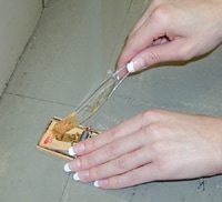
person baiting a snap trap with peanut butter

Trap Up!

Trap rodents around the home to help reduce the population.

various food containers with properly sealed lids

Clean Up!

Avoid illness: Take precautions before and while cleaning rodent-infested areas.


And for more information, the CDC offers a   16 page PDF  on Hantavirus, which is available on their Hantavirus Main page.


image

»» Read More